


FollowMyHealth™ Patient Portal
Communicate with your health care team
Patient Billing
Pay your bill or get answers to your billing questions
Our Specialists
Find out what we offer
Find a Provider
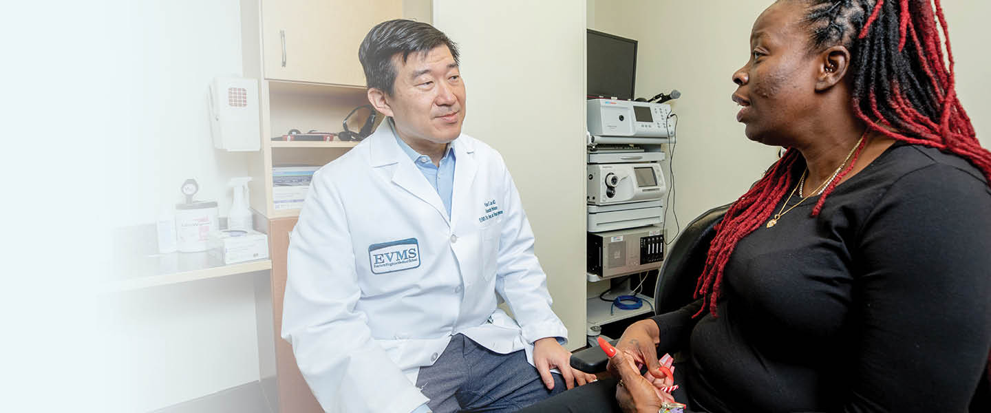
EVMS Medical Group clinical practices offer up-to-date, advanced, patient-centered care.
The EVMS Difference
As an academic medical center, EVMS offers the most advanced treatment options, comprehensive services, award-winning medical research and specialized patient-care programs. We continuously collaborate with national and international partners to develop medical technologies for the future. Choose providers with the knowledge to treat you better at EVMS Medical Group.